Need help with ECG interpretation

Nursing Students SRNA

Published

Questions are:

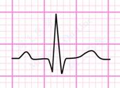
(1) P-wave - what EXACTLY causes the down slope of the P-wave? I understand the P-wave is atrial depolarization, so UPWARD portion/slope of the P-wave makes sense, BUT if depolarization is still occurring during the P-wave, WHY would the slope then go DOWNWARD? I would think that (a voltage drop as indicated by the downward slope on the 2nd half of the P-wave) indicates REpolarization (ie voltage drop), BUT repolarization of the atrium doesn't occur until later during QRS (masked by depolarization of the ventricles). Basically, it seems there should be NO downward slope on P.

(2) QRS - same question: WHY the downward slope (on the second half) if in DEpolarization? (ie since repolarization of vetricles doesn't occur until T)

(3) why the UPward slope on the T-wave if T-wave is REpolarization? (Would think REpolarization would be DOWNward since voltage difference gets greater/ie drops again during repolarization)

Thanks in advance

Specializes in ER.

What goes up must come down, in this case the "coming down" is the ECG tracing returning to baseline after the depolarization of the atrial tissue. The initial electrical impulse from atrial tissue causes a deflection in the ECG tracing as the electrical impulse travels from the atria at the SA node toward the AV node (the upward slope of the P wave) after which the tracing returns to baseline (the downward slope of the p wave). Same basic physiology with the other waves. This link may help: http://webtools.delmarlearning.com/sample_chapters/0766805204_02.pdf

This ->myocardactionpotential.gif

is different from this ->image004.png

The first is the voltage difference between inside and outside the cell. That's what your thinking about with up being depolarizing, and down being repolarizing.

I can't recall exactly how best to describe an ecg tracing (plus just got off shift), so correct me if I'm off. But think of it less as the charge and more like you've got the two leads (e.g. RA and LL) one is negative, one positive. When the current is moving towards the positive, the tracing goes up, and down towards the negative.

Uh, hmmmm. I think you are missing some basics that would help you tie the whole thing together.

I'm just not sure how to clear it up for you. You may need to go back to some of your A&P, and clear up some of the basics of cellular (muscle) electrical workings as this is key. All of it is tied together.

Yes, go pop your A&P book for a bit.

Specializes in ER.

What goes up must come down. In this case, the depolarization of the atria cause the upward deflection of the ECG tracing ("upward slope") and then the tracing returns to baseline (downward slope) when the electrical impulse has been conducted through the atria. Same basic concept with the QRS. See this link for more information about the electrophysiology of the heart: http://webtools.delmarlearning.com/sample_chapters/0766805204_02.pdf

Specializes in Critical Care.

In a nutshell, waveforms above the isoelectric line indicate that the net flow of electricity is coming toward the point of reference (the electrode) while waveforms below the isoelectric waveform indicate the net flow of electricity is heading away from the point of reference.

Specializes in Critical Care.

The direction of the wave has nothing to do with it being depolarization or repolarization. It has to do with the directional flow of the eletrical impulse, and the strength of that impulse.

Specializes in Critical Care, Education.

The direction (up, down, iso) is relative to the lead you're examining. If the current (valence) is moving toward the lead, it will cause an upward movement; if it is moving away from that lead, it will cause a downward movement. So, when you are monitoring in Lead II, the p wave, QRS & T wave are normally upright - simply because of where Lead II is located. In a 'normal' 12-lead, some QRS's are normally inverted due to where they are in relation to the way a normal heart depolarizes.

If you see alterations in the normal waveforms, it means that something has happened to change the normal current pathway. Figuring out what that 'something' could be? That will require more information, but once you understand it, you can apply it each and every time... for instance, under normal circumstances, elevated ST in leads II, III & AVI indicate a problem with oxygenation in the inferior area of the heart.

Hang in there - I found that it usually took 3 EKG courses for everything to actually click for most students unless they are able to work with an experienced preceptor in an environment with continuous cardiac monitoring.

Specializes in ICU + Infection Prevention.

It's a measure of current flow and direction of flow relative to the leads.

It is a hard value on cell polarization.

Have you taken any physics, because that will make what I am about to state easier to understand?

When you look at an ECG, you essentially have two components; magnitude and direction. So, we are talking about depolarisation with magnitude that takes a certain vector. The vector is in respect to the lead that you are looking through.

With the P wave, the positive deflection indicates that you do have a movement of ions and such a change in the potential of the cardiac cells. The positive deflection also indicates that you are observing this process from a negative to positive lead (view). Since a normal P wave does not go below the baseline or invert (assuming lead II), you are looking at depolarisation in a negative to positive vector (if we define our vector as movement from negative to positive) if you will.

So, the downward slope of the P wave is not telling us the vector is changing, otherwise it would fully invert and go downward (invert). So, really all it tells us, is there is a change in magnitude. In that, I mean, the atrial cells are all nearly depolarised. So, with fewer cells left to depolarise, we have decreased magnitude measured.

The QRS complex is somewhat similar, but now we do note that the Q wave and part of the S wave has negative deflection. The Q wave represents depolarisation of the septum from left to right. Since, the vector changes from what the lead is looking at, it is seen as negative deflection. Then, the ventricles depolarise, resulting in the large R wave, and finally, the base of the left ventricle depolarises, leading to the S wave.

Hopefully, you now understand why the T wave is upright. The ECG does not care that different electrolytes are moving, the only thin it measures is magnitude and direction. Since the direction of repolarisation is the same as seen through lead II as most of depolarisation, the T wave will be upright.

Specializes in Critical Care/Coronary Care Unit,.

I'm an ICU nurse..but that's still a little too technical for me. I just know how to interpret it and how to treat it. Your best bet would be to ask an EPS doctor or look on an EPS website.

Specializes in ICU + Infection Prevention.
It's a measure of current flow and direction of flow relative to the leads.

It is a hard value on cell polarization.

Due to moderation, I wasn't able to edit. That should read:

It's a measure of current flow and direction of flow relative to the leads.

It is an absolute value on cell polarization state.

Thus once a repolarization or depolarization reaches its maximum rate (peak of the P or T), the rate will then slow until no re/depolarization is occurring (the trace will decrease in amplitude back to the baseline). Whether the line goes up or down is entirely dependent on what the measured potential is on the current lead, that is, what direction is the current flowing relative to the electrode placements.

+ Add a Comment