- Community
-
Programs
- Schools
-
Careers
- RN Specialties
- Best RN Jobs and Salaries
- Aesthetic Nurse
- Nursing Informatics
- Nurse Case Manager
- NICU Nurse
- Forensic Nurse
- Labor and Delivery Nurse
- Psychiatric Nurse
- Pediatric Nurse
- Travel Nurse
- Telemetry Nurse
- Dermatology Nurse
- Nurse Practitioner
- Best NP Jobs and Salaries
- Family NP (FNP)
- Pediatric NP
- Neonatal NP
- Oncology NP
- Acute Care NP
- Aesthetic NP
- Women's Health NP
- Adult-Gerontology NP
- Orthopedic NP
- Emergency NP
- Psychiatric-Mental Health NP (PMHNP)
- APRN
- Nurse Educator
- Nurse Administrator
- Certified Nurse Midwife (CNM)
- Clinical Nurse Specialist (CNS)
- Certified Registered Nurse Anesthetist (CRNA)
- Resources
- Education

As a Cath Lab Nurse, I get to see a side of support devices up close and personal. One of my favorite assist devices is the Impella Heart Pump. This small pump made by ABioMed focuses on native heart recovery for patients. The pump allows the heart to rest and recover by off-loading the blood volume in the left ventricle into the ascending aorta. The pump accomplishes this by the Impella device that sits with one end in the left ventricle and the other end in the ascending aorta. In the left ventricle sits the inlet area on the device where blood flows into the device. Blood flows upward in the device by the power of the motor and redirects flow out into the ascending aorta. The motor creates negative pressure, thus off-loading the workload of the left ventricle to pump blood forward. The left ventricle can still pump blood on its own however, the Impella device is meant to do most of the work.
At maximum flow, the Impella heart pump can assist in providing 5.5 liters of blood per minute. Overall, the pump improves cardiac output (stroke volume x heart rate) by improving stroke volume.
The Impella device can be used for various situations and is approved for use by the FDA. The most common including cardiogenic shock, and protected PCI (percutaneous coronary intervention, aka stenting). In cardiogenic shock, the heart is unable to pump effectively, causing low blood flow back to the coronary arteries and systemic circulation. In a protected PCI, a patient who is not a candidate for CABG (coronary artery bypass graft) surgery, chooses not to get a CABG, is not recommended for surgery, or is high risk, has the Impella device inserted in before coronary stents are placed for heart protection. When a stent is deployed in a coronary artery, the stent balloon temporarily cuts off all blood supply to the myocardium distal to the stent balloon and can cause stress to the heart. In some cases, the heart can become stunned or stressed enough to the point that the patient can go into life-threatening dysrhythmias such as Vfib or Vtach. Due to this high risk, the Impella device provides constant blood flow back to the heart and systemic circulation EVEN during Vfib and Vtach. The patient would still need to be shocked per ACLS protocol to stop the dysrhythmia. I have heard of a patient on my previous unit that had the Impella device in, and went into Vfib, but was alert and talkative! The Impella was providing his cardiac output to perfuse his brain and the rest of his body.
In the Cath Lab, the patient is prepped and given conscious sedation to keep the patient comfortable and provide pain relief. Depending on the version of the pump that is used (there are 5; 4 for the left ventricle and 1 for the right ventricle), the insertion site is chosen, and a small sheath (usually the standard 6-7 French size) is inserted into the femoral or subclavian artery. At my facility, we commonly use the right femoral artery and the Impella CP (cardiac power) with smart assist version. This article will reflect information solely for the Impella CP with smart assist version. When the controller is set up, it is primed with either dextrose in 5% water, or a solution containing heparin. The dextrose is always used to prime the controller when initially setting up the controller and inserting the Impella in the procedure. The dextrose is then switched out when the patient arrives back to the unit with the Heparin-containing solution to keep the sheath free of clots and keep the patient anticoagulated. During the procedure, heparin IV push is given and ACTs are frequently checked to ensure the patient's ACT is above 250 so no clots form in the sheath. The FDA has only approved those two IV solutions which is why those two are only used.
During the procedure, the sheath size is dilated to a 14 French (the size of a pencil diameter) in the artery, and the device is inserted up into the aorta. The device is pushed further up and down into the heart, passing down through the aortic valve, to its home base with one end in the left ventricle, and the other end above the aortic valve in the ascending aorta. Interventional cardiologists verify placement with fluoroscopy and by assuring the radiopaque marker (a black marker seen on fluoroscopy) is sitting on the annulus (most distal part of the aortic valve). Once placement is verified, the Impella controller (brain) is turned on, and the motor begins to run. Everything is sutured into place and the patient is taken to the ICU for management. ABioMed has representatives that are alerted as soon as an Impella controller is turned on, and they come to the lab as soon as possible (someone is always on call). They offer expert guidance to the Cath lab team and the care team of the patient back on the unit. The reps can see a patient's Impella controller screen in real time from their work phones for real-time management and device support and are available 24/7!
Impella Control Screen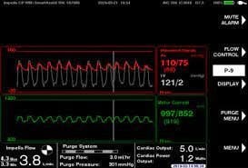
The most crucial tool is the Impella control screen. The screen provides specific information that reveals support, positioning, native (patient) cardiac output, fluid status, and how the device is working. Let's get started!
I personally was easily confused on what each section meant, so I turned to an ABioMed representative that frequently assists our Cath lab.
Red
The red box: the "AO" or aortic pressure and waveform shows an *estimated only* pressure; therefore, I put AO in parentheses. The "AO" pressure on the screen is NOT a true aortic blood pressure. Why? There is no transducer, therefore it cannot be zeroed to the atmosphere like a regular A-line and cannot be used for any clinical decision making or guidance at this time. The "AO" pressure can be as great at 10-15mmHg~ difference from the patient's true aortic pressure. Many facilities use the true blood pressure by using the cuff or have another A-line placed (radial or femoral). It is extremely important to note that the "AO" screen pressure is inaccurate because you do not want to intervene for a false low MAP or pressure, when in fact, the pressure isn't accurate and can be within normal range.
It is also important to note that the diastolic number is not negative. If the diastolic were to read a negative value, this should alert you of two things. First, the patient is dry and needs volume. With a swan in, we can assess CVP (central venous pressure). Remember, CVP is the pressure of blood in the right atrium and reflects fluid volume status (whether a patient is dry or overloaded). A normal CVP on average is between 4-12 mmHg. The second reason would be right ventricular failure. Assess how fluids help, and then if fluids make no change in increasing CVP, an echo might need to be done to see how well the right ventricle is pumping. Both situations are the most common cause for a suction alarm.
The "mean AO" pressure on the controller represents afterload if the patient does not have a swan in place for specific measurements of afterload (aka SVR/SVRI). Goal "AO map" is 60-80 mmHg. A "map" above 80 mmHg can represent an increased or elevated afterload, and that the patient might need afterload reduction (I.e., a Nipride gtt for example). Remember, the SVR/SVRI is always the most accurate indicator of true afterload value. Note that the Impella cannot give optimum support if afterload is too high. The greater the afterload, the more force is needed to push blood from the left ventricle into the aorta.
A very low "map" 30 mmHg or less with a suction alarm can indicate two situations. You need to start CPR, or the device is down too far and close to the aortic valve. You will know when your patient is coding! If the patient is stable, the device will need repositioning.
The "AO" pressure waveform should always be pulsatile as should every aortic waveform (we already know this).
Below the "AO" waveform you will see another waveform which is the left ventricular pressure. The left ventricular pressure is also estimated, as is the aortic pressure. The control screen shows both simultaneously to always show correct placement of the device. The left ventricular systolic peak of the waveform will line up exactly with the aortic systolic peak. We expect that both the left ventricle and aortic peaks will occur at the same time since the aortic valve is open is systole. There are sensors in the device in the LV and the ascending aorta which provide the waveforms and pressures.
Both peaks should always be in line with each other and both waveforms should always be touching one another. If both waveforms are apart from each other, this indicates that the device is against the heart muscle wall and needs to be repositioned.
Note- there will be no LV waveform present with a P-level 3 or less.
Green
The green box shows the motor waveform, and the waveform should always look pulsatile like an aortic waveform.
A flat motor waveform can indicate that the Impella device is entirely in the left ventricle or entirely up and in the aorta. Either way, the device will need to be repositioned.
Impella Flow
This area shows our maximum systolic and maximum diastolic Impella assisted cardiac output in liters/min. This number represents the cardiac output only being provided from the Impella. The largest number is the mean of the systolic and diastolic values.
Patient's native cardiac output
Cardiac output is the value you will get from your swan thermodilution or FICK calculation depending on provider preference. Select the "MENU" button, and then scroll down to "Enter cardiac output.” The value can be updated to reflect current patient data. Native cardiac output is calculated by subtracting the Impella flow value from the swan cardiac output value. When weaning, trends can be assessed by following native cardiac output as the Impella power flow (P value) is decreased, and less support is provided to the patient.
Cardiac power output
Cardiac power output is a value that represents the patient's mortality risk. An average goal is 1.0 and above. A value 0.8 or less is indicative that a provider needs to be aware. This value is calculated by multiplying cardiac output and map, divided by a constant. This value is also important to watch for trends. The lower the value, the higher the mortality risk. You will see other values change on the screen as this number changes, further representing there is a problem.
P-level
Last, but not least we have our P-level on the screen. The P-level indicates the power level of the motor while running. P-levels range from P-9 (maximum) to P-0 (controller turned off). When the controller is turned on for the first time for a patient, the controller will set the P-level to automatic, which is equivalent to P-9. A patient is weaned from support by decreasing P-level.
Code blue
If your patient requires CPR, always turn the P-level to P-2. Patient blue = P-2. This turns the motor down but not off. It is safe to perform CPR with the Impella device in use. After achieving ROSC, the P-level can be increased per provider order.
A representative is available 24/7, so if you have a simple question or need guidance with the controller, call the number listed on the controller. It is always safest to ask than assume and risk patient safety.
The Impella heart pump is an amazing device that protects a patient's native heart function. You can view real patient stories by going to AbioMed.com and click on "Explore patient stories.” You will be amazed! I have no affiliation with AbioMed or am receiving any profit from this company; I simply just love this device and what it has done for patients.
FACT: Remember, if it was easy, anyone could do it!
About BuckeyeICUNurse, BSN
Megan is a Registered Nurse in Ohio with experience in neonatal, cardiac, and pulmonary intensive care, as well as invasive cardiac cath lab.
Share this post
Share on other sites