confused about cardiac output....

Nursing Students Student Assist

Published

Hi everyone,

can someone please read the following and either tell me i'm correct or explain why I'm wrong?

Am I understanding the following concepts properly? I'm most confused about preload.

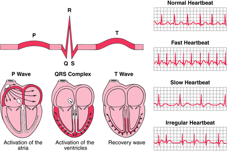
Heart rate: bradycardia can decrease cardiac output (if the heart isn't pumping fast enough then cardiac output can go down). Tachycardia can decrease cardiac output (if the heart is pumping too fast, the heart doesn't have time to fill and then cardiac output can go down). That's why you want the heart rate in the "sweet spot" of 60-100 bpm.

Afterload is the resistance the left ventricle has to fight to push the blood out, and is a combination of systemic ventricular pressure and aortic pressure. Increasing afterload would decrease cardiac output, correct? So vasodilation decreases afterload, correct?

Contractictility of the heart is how strong/intensity of the contraction is...you want a strong contraction to push more blood to the body so increasing contractility of the heart (done with inotrope medications) increases cardiac output, correct?

How does preload factor in to this? I've heard people refer to preload as stretch....I don't totally get that. My understanding is that preload is the volume of blood in the right ventricle at the end of diastole....meaning it is the volume of blood that gets "left behind" in the heart and doesn't get to the body where it needs to be....meaning that decreasing preload would increase cardiac output? Is that correct? How would preload ever get increased? would we ever want preload increased?

Your help is much appreciated!

Specializes in Critical Care, Education.

You're almost there!!

Pre-load is a term that can be applied to either chamber. It is easier to understand if you separate the sides of the heart. Remember that the Right ventricle has to push through the lungs. Increased pulmonary resistance will INCREASE RV afterload.... usually causing blood to back up into the RV & RA - which causes excessive pre-load if the RV begins to decompensate. Excessive intravascular fluid volume (due to any cause), pulmonary disease and valve problems are most commonly associated with this. If it's chronic, you'll probably see atrial arrhythmias due to a stretched out (irritable) RA. This is often the first obvious sign of R heart overload.

Also, remember Starling's law - in a normal heart, the heart will eject a greater stroke volume if it is filled to a greater volume at the end of diastole (pre-load). However, this is dependent upon ventricular oomph as well as afterload. Diseased heart muscle can be floppy or rigid... both ends of the spectrum will decrease contractility. If the muscle is too stretched, it does not 'snap back' as it should. So, at a certain point, excessive pre-load will cause too much stretching. The amount of blood ejected decreases because the muscle is not capable of squeezing hard enough to empty itself and at that point, retro-grade volume build up starts to happen.

Physically, you'll notice wet lungs beginning at the bases & working it's way up as the condition worsens. You'll notice venous engorgement in the neck & increasing central pressures (CVP, PWP, etc) Respiratory rate will increase.... Atrial arrhythmias and so on. So - there are many occasions when decreasing pre-load is a therapeutic goal.

On the flip side - You may be working to increase pre-load in any situation in which there is insufficient intravascular fluid (dehydration, bleeding, 3rd spacing, etc) in order to maintain adequate cardiac output. This is when you'll get orders to rapidly infuse fluids & volume expanders.

Isn't this fun???

How does preload factor in to this? I've heard people refer to preload as stretch....I don't totally get that. My understanding is that preload is the volume of blood in the right ventricle at the end of diastole....meaning it is the volume of blood that gets "left behind" in the heart and doesn't get to the body where it needs to be....meaning that decreasing preload would increase cardiac output? Is that correct? How would preload ever get increased? would we ever want preload increased?

Pretty close, but missing one key idea. The end of diastole is just before the beginning of systole, so there's nothing related to "left behind."

Let's start at the beginning and see if I can help you understand this a bit better.

Filling pressure is just the pressure that is in the ventricles at the end of diastole. For a given volume delivered to a ventricle, pressure can be lower if the ventricle is nice and soft and flexible and empty, ready to accept a new load, than if it's hard and scarred up or has leftover blood in it from the last systole because the AV is hard to open OR because its contractility was so lousy that it didn't empty well. Another term that is used could be "preload," pre- meaning "before systole," and load, well, being the load of blood delivered to the ventricle that it is gonna have to move out in systole. You can measure load as weight or volume, but the way we look at it is by measuring the pressure that occurs there. Pressure changes tell us what's going on in there. Think about a balloon: If it's soft (low pressure), is there more or less air in it than when it's hard (high pressure)? That's why pressure tells you about volumes.

First, let's look at the blood flow in a linear fashion.

Body > Veins > Vena Cava > Right Atrium > tricuspid valve > Right Ventricle > pulmonic valve > Pulmonary Artery > LUNGS >Pulmonary Vein > Left Atrium > mitral valve > Left ventricle > aortic valve > Arteries > Body

Think about when the valves between two chambers are OPEN. By definition, each chamber must be at the same pressure, right? So, at the end of diastole, just before systole, the pressure in the LV is the same as LA pressure is the same as the pressure in the pulmonary vein (no valve in the way there) and in the pulmonary capillary bed. And since there are no valves in the pulmonary capillary bed, tracking backwards, you can see that LV end diastolic pressure equals end-diastolic PULMONARY ARTERY PRESSURE, which is, conveniently, what we look at when we are wondering what's going on in the left heart.

OK. Now, why do we care about LV end-diastolic (filling) pressure (PRELOAD)? For that, I wish I could draw you a nice little curve here. I am going to try to attach it, but if it doesn't show up, I can't, so I will describe it and YOU will draw it on a piece of paper to look at while we chat.

Oh, it worked!

Horizontal axis: label this "preload" or any other term you like. Filling pressure, PA diastolic pressure is the same thing (see above) and you can even extrapolate all the way back to central venous pressure, for a rough trend-setting bit of data.

The vertical axis you will call "cardiac output," or "blood pressure," or "stroke volume," like the illustration, because the line we are going to draw is going to explain something really cool.

Start lowish on the left, near the vertical axis-- low filling pressure means low BP. Think: hemorrhage, hypovolemia, makes your BP low, right?

Slant the line upwards to the right, showing that blood pressure (cardiac output) increases the more blood you put into the heart. (Tank up that hypovolemic guy, and BP improves.) But at some point, that upward-going curve peaks, flattens out...and then it DROPS as the preload keeps increasing. This is because cardiac muscle is like a rubber band-- the more you stretch it, the harder it contracts...to a point, at which point it gets too stretched out and actually contracts less well. Draw a little asterisk at the top of that curve, where it starts to fall, then let it fall a little bit. That asterisk marks the best cardiac output you can get-- preload and output are optimal for that heart. Beyond that point, where the line slopes downwards, lies congestive heart failure- the heart is too full, has more than it can handle, and it fails. (This is, BTW, called the Frank-Starling Law of the heart, and you just drew the Frank-Starling curve) Pressure backs up into the pulmonary capillary bed making the lungs get wet and heavy. This is when people get diuretics (to decrease that excessive preload) AND drugs to improve their contractility.

Of course, if contractility is lousy because of coronary artery disease, previous MI, or whatever, this whole curvy line thing will kinda slide over to the left-- the myocardium will fail with lower pressures than it would if it had better contractility. Better contractility (a right shift) means it will handle more preload (higher filling pressures) and make better BP out of it. Draw a second curve to the right of the first one, parallel to it, to see that.

(In the illustration, you have two lower curves, one labeled "HF," for "heart failure," and one showing HF with treatment-- better inotropic support, better oxygenation to the coronary arteries, lower afterload, all of the above, whatever makes it contract better.) With me so far?

I think you can see how CAD will give you higher filling pressures-- when the heart is failing a bit, it goes past the top of its curve more easily because its contractility is diminished.

Mitral STENOSIS will, in fact, decrease your LV preload, but it will increase pressures back into the lungs and, eventually, the right heart, because of the resistance to flow from the right side to the LV. Mitral REGURGITATION, on the other hand, will result in higher filling pressures because when the ventricle contracts in systole, some of the blood goes backwards, leaving excess sloshing around between the atrium and ventricle; the ventricle will have to accept a higher reload at diastole, and it doesn't like it. Over the top of the curve again.

Well, I hope this hasn't confused you. I used to tell my students they had to know this because we saw lots of people with all sorts of deficits, but if they didn't have hearts and lungs, they were dead and we didn't have to take care of them anymore. Works in every possible area you could work, except pathology. Please ask me if I've confused you anywhere.

thank you both, and sorry for the delay in responding.

I now understand that it is not an absolute...at times you want the preload increased to increase cardiac output (example, when someone's blood volume is very low you want to increase their preload...increasing their blood volume will increase their preload.) at other times when their preload is too high and the heart muscle has too much stretch (like when the heart is fluid overloaded, you give hte patient diuretics in order to decrease the preload and increase the cardiac output).

thank you! I am sorry for the delayed response, and am very grateful.

One more thing to remember-- you want to give someone in failure inotropes, to increase contractility, IF doing so doesn't increase the oxygen demand more than his lousy coronary arteries can keep up with.

Someone who is hypovolemic is already doing his best on the contractility front, and will do more when he gets a better preload, so you DON'T want to whip his heart harder with inotropes.

thanks. to clarify

decrease cardiac ouput related blood loss - low bp high pulse restlessness hypovolemic shock or heart failure

decrease peripheral circulation related hypoxia cyanosis

decrease blood vessel related to dec heart muscle elasticity - dvt - pvd

question so does claudication both related to peripheral circulation and/or decrease venous return?

thanks. to clarify

decrease cardiac ouput related blood loss - low bp high pulse restlessness hypovolemic shock or heart failure

It's not the heart's fault if it's not getting enough blood to pump, so it's not heart failure... yet. Cardiac failure occurs if/when the hypovolemia is so bad that there is inadequate oxygen being delivered to the heart.

decrease peripheral circulation related hypoxia cyanosis

You're kinda losing me here. Punctuation is your friend-- please do not abandon it so heartlessly! :)

Do you mean that decreased peripheral circulation is caused by / related to hypoxia and/or cyanosis? The answer there is no. These things are effects of inadequate perfusion (blood flow), although cyanosis will be a very late sign. Lots of people are seriously hypovolemic with low BP and not turning grey-blue.

A limb without adequate arterial blood flow can become cyanotic, or at least dusky. Not usually what you see with hypovolemia, but you do see it with peripheral arterial disease (PVD, obliterans, or arterial embolus cutting off blood flow into the limb).

Can you clarify what you were thinking here so I can work with it?

decrease blood vessel related to dec heart muscle elasticity - dvt - pvd

OK, I'm pretty lost here. What do you mean by "decrease blood vessel" and related to/caused by "decreased heart muscle elasticity"? Do you mean "contractility," the ability of the heart to contract to move blood along, or are you thinking about "elasticity," its actual level of stiffness?

And where do deep vein thrombosis (clots in deep veins) and peripheral vascular disease (generally accepted as arterial) fit in? Help me out; tell me what your thought process is so I can help you.

question so does claudication both related to peripheral circulation and/or decrease venous return?

Are you asking, "Does claudication related to peripheral circulation and/or decrease venous return ..." do what? I have a hard time figuring out what you are asking.

Claudication is pain, usually in muscles, that occurs because they aren't getting enough blood flow due to peripheral arterial disease. So yes, claudication is related to bad peripheral perfusion (circulation). It isn't caused by hypovolemia, though, or decreased venous return. People with claudication have pain when they walk because their exercising muscles aren't getting enough oxygen, usually relieved by sitting and resting. They also usually have other signs of poor arterial flow, like thickened nails, loss of hair in the limb, poor or absent peripheral pulses, and cool skin below the level of the obstruction.

The major problem with deep vein thrombosis (clot in the vein) isn't decreased venous return, because other veins will mostly take up the slack. It's the risk of that thrombus (clot) will break loose and travel to the lungs (pulmonary embolus), a whole 'nother can of worms.

Am I getting closer to your misconceptions? Please tell us what your ideas are in complete sentences :) so we don't have to guess.

Specializes in Critical Care, ED, Cath lab, CTPAC,Trauma.

preload = pressure inside the heart during diastole (resting phase)

-starling curve. to a point, the more volume sitting in a chamber, the better a heart works

afterload = pressure the heart has to overcome to squirt blood out of the ventricle

-anything that causes resistance. from a malfunctioning valve to tight arteries to standing on your head.

Gross analogy, but works for me....

Thing about flushing a toilet......

you flush, and then flush again right away......nothing happens right? This is because the tank doesn't have time to fill....PRELOAD is decreased in the tank.

What if the toilet is plugged up? When you flush, it backs up....this is too much afterload.

Specializes in Critical Care, ED, Cath lab, CTPAC,Trauma.

Try here......https://allnurses.com/nursing-student-assistance/preload-vs-afterload-887678.html

Preload is stretch. The amount of volume being returned to the right side of the heart from systemic circulation.

Afterload is squeeze. The amount of resistance the left side of the heart has to overcome in order to eject blood.

The right side of the heart, composed of the right atrium and ventricle, collects and pumps blood to the lungs through the pulmonary arteries. The lungs refresh the blood with a new supply of oxygen, making it turn red.

Oxygen-rich blood, "red blood," then enters the left side of the heart, composed of the left atrium and ventricle, and is pumped through the aorta to the body to supply tissues with oxygen.

Four valves within your heart keep your blood moving the right way. The tricuspid, mitral, pulmonary and aortic valves work like gates on a fence. They open only one way and only when pushed on. Each valve opens and closes once per heartbeat — or about once every second.

A beating heart contracts and relaxes. Contraction is called systole, and relaxing is called diastole.

During systole, the ventricles contract, forcing blood into the vessels going to your lungs and body — much like ketchup being forced out of a squeeze bottle. The right ventricle contracts a little bit before the left ventricle does. The ventricles then relax during diastole and are filled with blood coming from the upper chambers, the left atria (contains oxygenated blood from the lungs/luminary vein)and right atria (from the Superior/inferior vena cava). Then the cycle starts over again.

The heart is nourished by blood vessels called coronary arteries extend over the surface of your heart and branch into smaller capillaries. The heart also has electrical wiring, which keeps it beating. Electrical impulses begin high in the right atrium and travel through specialized pathways to the ventricles, delivering the signal to pump. (PQRST) The conduction system keeps the heart beating in a coordinated and normal rhythm, which in turn keeps blood circulating. The continuous exchange of oxygen-rich blood with oxygen-poor blood is what keeps us alive.

tumblr_lzjrhzv5Fh1qc7n4go1_500.gif

404 Not Found

ECG Learning Center - An introduction to clinical electrocardiography

ECGpedia

images?q=tbn:ANd9GcRksBpnpPIZpT7BuWKSU4O4O9-8qbNhC2E-1UKt_R_lXVBxvR0c

newreply.php?do=postreply&t=979164

Preload is about volume. If the water pressure is low, the water output will be a trickle, not enough to water and sustain your pretty garden. If the volume is too much, it will back up your plumbing system (Right-sided heart failure engorged liver, systemic edema, etc, or left-sided failure pulmonary edema.)

Afterload is about pressure or resistance. If there is a link or narrowing in your garden hose, the volume will back up AND the output will drop. Think of it like a garden hose.........

A good analogy is that your hose has gone form a garden hose to a fire hose or developed huge leaks and it will either get more blood to the right places easier (decreasing afterload so the heart doesn't have to work as hard and will therefore work better) ...or you will not get the blood to where it need to go until you "patch it up" (constrict the blood vessels to a more normal size like the vasodilation that occurs with sepsis)or stop the bleeding by sealing the leak(the source of hemorrhage)....OR....

Gross analogy, but it works...Think about flushing a toilet......you flush, and then flush again right away......nothing happens right? This is because the tank doesn't have time to fill....PRELOAD is decreased in the tank.

What if the toilet is plugged up? When you flush, it backs up....this is too much afterload.

Preload = Volume

Afterload = Pressure/Resistance

404 Not Found Pressure/BP010.htm

good to know it GRN. Your explanation is best. I have a question how can the ARB help to reduce the afterload?

good to know it GRN. Your explanation is best. I have a question how can the ARB help to reduce the afterload?

OK, you can figure this out yourself. What's the A-- angiotensin, right? What does it do and where?

What's a R-- receptor? So there are receptors for angiotensin. Hmmm. Where are they, and what happens when a little angiotensin wanders by them?

and B -- blocker. Ahh, there are blockers for those angiotensin receptors. Then what happens-- or more to the point, what DOESN"T happen when that angiotensin wanders on by?

Now go back and look at what afterload is and tell me the answer to your question.

HINT but no fair looking until you give it some thought and look in your pharmacolgy book for the answer to my questions: http://www.medicinenet.com/angiotensin_ii_receptor_blockers/article.htm

+ Add a Comment