- Community
-
Programs
- Schools
-
Careers
- RN Specialties
- Best RN Jobs and Salaries
- Aesthetic Nurse
- Nursing Informatics
- Nurse Case Manager
- NICU Nurse
- Forensic Nurse
- Labor and Delivery Nurse
- Psychiatric Nurse
- Pediatric Nurse
- Travel Nurse
- Telemetry Nurse
- Dermatology Nurse
- Nurse Practitioner
- Best NP Jobs and Salaries
- Family NP (FNP)
- Pediatric NP
- Neonatal NP
- Oncology NP
- Acute Care NP
- Aesthetic NP
- Women's Health NP
- Adult-Gerontology NP
- Orthopedic NP
- Emergency NP
- Psychiatric-Mental Health NP (PMHNP)
- APRN
- Nurse Educator
- Nurse Administrator
- Certified Nurse Midwife (CNM)
- Clinical Nurse Specialist (CNS)
- Certified Registered Nurse Anesthetist (CRNA)
- Resources
- Education


Chrystal Marie
4 Posts
Hello all :) I am new to the forum and am in need of some help with my care plan for this term. I am currently doing a clinical rotation on a telemetry floor (so cool!) but unfortunately, we have only been learning about the diseases of the heart for a short period of time. Here is the information that I have about my patient:
Patient is a 95 yo female admitted with possible acute coronary syndrome; chest pain
(Patient presented to the ER after an episode of altered mental status. The staff at the assisted living facility where she resides stated the patient did not recognize where she was or staff members. After admission to the ER, the patient had a bowel movement followed up by "stabbing" chest pain radiating to her entire upper back/shoulder area)
Vitals: T-98.4, P- 84, R- 14, BP- 148/70, Spo2%= 94% on room air
History: CAD, anemia, HTN, heart attack, cardiac shunt (to name a few)
Labs: RBC's- 3.48, Hgb- 10.7, Hct- 32.0, MCHC- 33.3
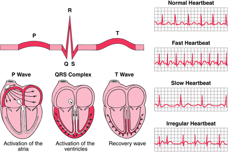
Assessment data: murmur (which was confirmed by cardiologist as an aortic stenosis murmur), monitor reading shows a prolonged PR interval of 0.24 (which I know is indicative of first degree AV block), absent pedal pulse on L foot, R foot 2+, no other issues with any other pulses- did a Doppler on L foot and still had no luck finding a pulse (neither did my instructor)... but extremities were warm and dry... pt did not complain of sob and was not having any current chest pain.
For this care plan we have to have a primary and a secondary diagnosis. For my primary I would like to do decreased cardiac output but am having some difficultly with the "related to" section because I am not 100% understanding of some of these disease processes. For secondary I would like to do risk for falls. I chose this d/t her age, also she gets around strictly by wheelchair, has a hx of arthritis, osteoporosis, joint limitations, gait disturbance, and has had a L knee replacement, R hip replacement and L femur repair.
Any input is greatly appreciated (as well as criticism... I am student so be nice lol). I know it is hard sometimes for people to comment on care plans because they may not have enough assessment data, etc.... so if added information is needed please let me know.