Need help with cardiac system please

Nursing Students Pre-Nursing

Published

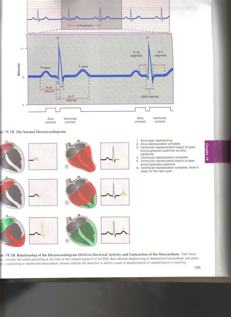
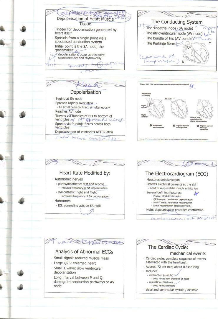
We are studying the cardiac system in phys and I am needing any and all websites that deal with cardiac conduction.... especially learning the ECG.

Any websites that have to do with reading EKG's would be so helpful. Our text is "okay" but I would like to view anything online to help me with this.

I've had the flu/cold for 5 days now and I know that part of my not "getting it" really well is due to that. Tomorrow aftgernoon (Tues) we have lab with ECG's and I need to understand it better.

The depolarization, repolarization thing is not clicking....... help!:uhoh3:

Thanks!!

MaryRose

Here are some of the notes I have on this topic:

Explanation of figure 19.16:

Depolarization/repolarization.

1) P wave is produced when a signal from the SA node spreads through to the atria and depolarizes them. Atrial systole begins about 100 msec after the P wave begins, during the P-Q segment. It represents the time required for impulses to travel from the SA node to the AV node.

2) The QRS complex consists of a small downward deflection (S). It marks the firing of the AV node and onset of ventricular depolarization.

(the complex shape of QRS is d/t different sizes of the two ventricles and different times req'd for them to depolarize)

Ventricular systole soon after the QRS complex in the S-T segment. Atrial repolarization and diastole also occur during the QRS interval, atrial repolarization sends a relatively weak signal that is obscured by the electrical activity of the more muscular ventricles. The ST segment corresponds to the plateau in the myocardial action potential and thus represents a time during which the ventricles contract and eject blood.

3) The T wave is generated by ventricular repolarization immediately before diastole. The ventricles take longer to repolarize than to depolarise; The T wave is therefore smaller and more spread out than the QRS complex.

http://img.photobucket.com/albums/v111/blue_eyes3/deprepol.jpg - typical ECG & diagram explain depolarization/repolarization, from Anatomy & Physiology by Saladin. K.

http://img.photobucket.com/albums/v111/blue_eyes3/lectureheart.jpg - lecture notes on depolarization/repolarization + ECH

It's a tough topic, but with a lot of studying you'll soon come to understand it. Hope this helps.

Specializes in MICU for 4 years, now PICU for 3 years!.

Maybe try to get a book specifically on EKG's... we just had an EKG test, and the book we used to study out of is "EKG Plain and Simple: From Rhythm Strips to 12-Leads" by Karen M. Ellis. It was about $50, but IMO, so worth it. It has about 6 chapters all devoted to the cardiac cycle, then it goes into how to read EKG's, and it gives you about 300 strips to practice on. Here at my school, we not only use it this quarter, but in two quarters it a required book again, maybe they have a book like that for students farther into the nursing program at your school.

Here are some of the notes I have on this topic:

Explanation of figure 19.16:

Depolarization/repolarization.

1) P wave is produced when a signal from the SA node spreads through to the atria and depolarizes them. Atrial systole begins about 100 msec after the P wave begins, during the P-Q segment. It represents the time required for impulses to travel from the SA node to the AV node.

2) The QRS complex consists of a small downward deflection (S). It marks the firing of the AV node and onset of ventricular depolarization.

(the complex shape of QRS is d/t different sizes of the two ventricles and different times req'd for them to depolarize)

Ventricular systole soon after the QRS complex in the S-T segment. Atrial repolarization and diastole also occur during the QRS interval, atrial repolarization sends a relatively weak signal that is obscured by the electrical activity of the more muscular ventricles. The ST segment corresponds to the plateau in the myocardial action potential and thus represents a time during which the ventricles contract and eject blood.

3) The T wave is generated by ventricular repolarization immediately before diastole. The ventricles take longer to repolarize than to depolarise; The T wave is therefore smaller and more spread out than the QRS complex.

http://img.photobucket.com/albums/v111/blue_eyes3/deprepol.jpg - typical ECG & diagram explain depolarization/repolarization, from Anatomy & Physiology by Saladin. K.

http://img.photobucket.com/albums/v111/blue_eyes3/lectureheart.jpg - lecture notes on depolarization/repolarization + ECH

It's a tough topic, but with a lot of studying you'll soon come to understand it. Hope this helps.

Thanks so much for the info and especially the photos! The photos made it all just "click" in my head a lot better.

I'm going to print that out and take to lab today!:)

Hugs!

MaryRose

Maybe try to get a book specifically on EKG's... we just had an EKG test, and the book we used to study out of is "EKG Plain and Simple: From Rhythm Strips to 12-Leads" by Karen M. Ellis. It was about $50, but IMO, so worth it. It has about 6 chapters all devoted to the cardiac cycle, then it goes into how to read EKG's, and it gives you about 300 strips to practice on. Here at my school, we not only use it this quarter, but in two quarters it a required book again, maybe they have a book like that for students farther into the nursing program at your school.

Thanks for the book info. I will look into this one for sure. I realize how vital it is to understand this info. This will be something for me to dig into while waiting to actually begin the nursing program.

I think for thisd class, getting past this flubug will be the biggest help right now. I'm still so foggy brainwise!

Hugs!

MaryRose

Specializes in Emergency Dept, M/S.

Thank you for the info!!

We are doing the Cardiac Unit now also. In fact, I'm writing and re-writing info on the cardiac cycle now (okay, I'm on a small break! LOL).

Whew.... made it thru lab without looking tooo stupid! lol

We got to run 2 lead ECG's on each other... that was fun. I thought we would have to know more than I anticipated.... so it was better than I expected.

We also took each other's BP, pulse etc.... no biggie. Luckily my buddie and I brought our own steth and BP cuffs in so we didn't have to wait in line which gave us more time to try out the ECG systems.

I am studying the cardiac conduction system this whole weekend!

Hugs!

MaryRose

+ Add a Comment